Running diagnostics

Running diagnostics
Showing posts with label Trauma. Show all posts

Thursday, February 22, 2018

Altered Level Of Consciousness

Today's post is also available in podcast form; visit www.medonthego.podbean.com to stream or download!

If you haven't already done so, also check out our Facebook page www.facebook.com/drolimedonthego --it has some hilarious medicine-related jokes and some useful, applicable articles too. If you wish to support us, please see www.patron.podbean.com/medonthego for more details!

Altered mental status is a collective, non-specific term referring to change in cognitive function, behavior, or attentiveness. It includes delirium, dementia, lethargy (state of decreased awareness and alertness i.e. patient may appear wakeful), stupor (unresponsive but rousable), and coma (sleep-like state, not rousable to consciousness).

Possible causes of coma (AEIOU TIPS)
Acidosis/alcohol
Epilepsy
Infection
Oxygen (hypoxia)/opiates
Uremia
Temperature/trauma (esp. to head)
Insulin (too little or too much)
Psychogenic/poisoning
Stroke


Remember “GCS < 8 intubate!” Ability to protect airway is first priority.

History
·      Obtain collateral from family, friends, police, paramedics, old charts, or Medic Alert bracelets etc.
·      Onset and progression
o   Antecedent trauma, seizure activity, fever
o   Abrupt onset suggests CNS hemorrhage/ischemia or cardiac cause
o   Progression over hours to days suggests progressive CNS lesion or toxic/metabolic cause
·      Determine patient’s baseline level of consciousness
·      Past medical history, paying attention to similar episodes, overdose, or concurrent psychiatric disorders (e.g. depression).

Physical examination
·      Vital signs including temperature
·      Cardiac, respiratory, and abdominal exams
·      Complete neurological exam
o   Pupil size and reactivity
o   GCS

Investigations
·      Blood work—rapid blood sugar, CBC, electrolytes, creatinine, BUN, liver function test, glucose, serum osmolality, venous blood gas, coagulation studies, troponins
·      Imaging—CXR, CT head
·      Other tests—ECG, urine analysis, urine toxicology

Diagnosis
·      Administer appropriate universal antidotes
o   Thiamine 100mg IV if history of alcoholism or patient looks malnourished
o   One ampule D50W IV if low blood sugar
o   Naloxone 0.4~2mg IV or IM if opiate overdose is suspected
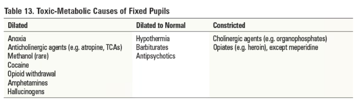
·      Distinguish between structural and toxic-metabolic coma
o   Structural coma
§  Pupils, extra-ocular movements, and motor findings are usually asymmetrical
§  Look for focal or lateralizing abnormalities
o   Toxic-metabolic coma
§  Dysfunction at lower levels of the brainstem (e.g. caloric unresponsiveness)
§  Respiratory depression in association with an intact upper brainstem (e.g. equal reactive pupils)
§  Extra-ocular movements and motor findings are symmetrical or absent
·      Essential to re-examine frequently because status can change rapidly
·      Diagnosis may become apparent only with the passage of time. Delayed deficit after head trauma suggestive of epidural hematoma (characteristic “lucid interval”).


Disposition
·      Admission if ongoing decreased level of consciousness

·      Discharge if altered level of consciousness is readily reversible; ensure adequate follow-up care

Friday, February 9, 2018

Genitourinary Tract Injuries

Welcome back to Med On The Go! This week we discuss genitourinary tract injuries commonly seen in the emergency room setting. Before we start, I'd like to invite you to visit our Facebook page www.facebook.com/drolimedonthego/ and help us by pressing "like". As usual, this blog post is also available in podcast form; subscribe to Med On The Go With Dr. Oli on iTunes, Google Play, or Podbean to make sure you never miss an episode. If you'd like to further support us, please visit www.patron.podbean.com/medonthego for more details.

Now let's dive into this week's topic:

Genitourinary tract injury secondary to trauma can occur any where within the genitourinary tract by a variety of mechanisms.
·      Blunt trauma—often associated with pelvic fractures
o   Upper tract
§  Renal
·      Contusions (minor injury—parenchymal ecchymoses with intact renal capsule)
·      Parenchymal tears/lacerations—non-communicating (hematoma), communicating (urine extravasation, hematuria)
§  Ureter: rare, at uretero-pelvic junction
o   Lower tract
§  Bladder—gross hematuria usually suggests bladder injury
·      Extraperitoneal rupture of bladder from pelvic fracture fragments
·      Intraperitoneal rupture of bladder from trauma and full bladder
§  Urethra
·      Posterior urethral injuries: MVCs, falls, pelvic fractures
·      Anterior urethral injuries: blunt trauma to perineum, straddle injuries/direct strike
o   External genitalia
·      Penetrating trauma—damage to kidney, bladder, ureter (rare), external genitalia
·      Acceleration/deceleration injury—renal pedicle injury—high mortality rate (laceration and thrombosis of renal artery, renal vein, and their branches).
·      Iatrogenic injury—ureter and urethra damage secondary to instrumentation

History
·      Mechanism of injury
·      Hematuria (microscopic or gross), any blood on underwear
·      Dysuria, urinary retention
·      History of hypotension

Physical examination
·      Abdominal pain, flank pain, CVA tenderness, upper quadrant mass, perineal lacerations
·      DRE: sphincter tone, position of prostate, presence of blood
·      Scrotum: ecchymoses, lacerations, testicular disruption, hematomas
·      Bimanual exam, speculum exam
·      Extraperitoneal bladder rupture: pelvic instability, suprapubic tenderness of mass of urine or extravasated blood
·      Intraperitoneal bladder rupture: acute abdomen
·      Urethral injury: perineal ecchymosis, scrotal hematoma, blood at penile meatus, high riding prostate, pelvic fractures

Investigations—in gross hematuria, the GU system is investigated from distal to proximal (i.e. urethrogram, cystogram… etc.)
·      Urethra: retrograde urethrography
·      Bladder: urinary analysis, CT scan, urethrogram +/- retrograde cystoscopy +/- cystogram (distended bladder and post-voiding)
·      Ureter: retrograde ureterogram
·      Renal: CT scan (best, if hemodynamically stable), intravenous pyelogram

Management
·      Urology consultation
·      Renal injury
o   Minor injuries—conservative management such as bed rest, hydration, analgesia, antibiotics
o   Major injuries—admit
§  Conservative management with frequent reassessments, serial urine analysis +/- re-imaging
§  Surgical repair (exploration, nephrectomy) for hemodynamically unstable patients or those who continue to bleed > 48hrs, major urine extravasation, renal pedicle injury, all penetrating wounds and major lacerations, infections, renal artery thrombosis
·      Ureter injury—ureterouretostomy
·      Bladder injury
o   Extraperitoneal
§  Minor rupture—Foley drainage for 10~14 days
§  Major rupture—surgical repair
o   Intraperitoneal—drain abdomen and surgical repair
·      Urethral injury
o   Anterior—conservative treatment, consider Foley catheter or suprapubic cystostomy and antibiotics for those who cannot void

o   Posterior—suprapubic cystostomy (AVOID CATHERIZATION) +/- surgical repair

Friday, January 19, 2018

Abdominal Trauma

As always, today's post is available in podcast form, available at www.medonthego.podbean.com. Subscribe on iTunes, Google Play, or Podbean to make sure you never miss an episode. Want to get to know us better? Check out our Facebook page at www.facebook.com/drolimedonthego/. Wanna support the podcast? See www.patron.podbean.com/medonthego.

Let’s open with a bit of trivia: what kind of injuries can you receive from a seatbelt?
A.     Retroperitoneal duodenal trauma
B.     Intraperitoneal bowel transection
C.     Mesenteric injury
D.    L-spine injury
E.     All of the above
If you chose D like me, you would be wrong! The answer is “all of the above”. Who knew seatbelts can cause so many abdominal injuries?!

Two main types of injuries can cause abdominal trauma: blunt (causing solid organ injury, with the spleen most commonly affected followed by the liver) and penetrating (causing hollow organ or liver injury). Penetrating injuries are most common.

Blunt trauma of the abdomen can result in two types of hemorrhage: intraperitoneal and retroperitoneal. When a patient presents with multi-system trauma, be extra suspicious of blunt trauma damage in the abdomen. A history focused on the mechanism of injury should be included in the general medical history.

Physical examination is often unreliable in multi-system trauma, with a wide spectrum of presentations; sometimes abdominal symptoms may be masked by other injuries—therefore serial examinations need to be done. Common findings include tachycardia, tachypnea, oliguria, febrile, and hypotension; signs of slow blood loss may not be apparent right away.
·      Inspect: contusions, abrasions, seatbelt sign, distention
·      Auscultate: bruits, bowel sounds
·      Palpate: tenderness, rebound tenderness, rigidity, guarding
·      DRE: rectal tone, blood, bone fragments, prostate position
·      Placement of NG tube and Foley catheter should be considered part of the abdominal exam. Foley catheter should be inserted in unconscious patient or patient with multiple injuries who cannot void spontaneously. NG tube can be used to decompress the stomach and proximal small bowel; but it’s contraindicated for patients with suspected facial fractures or basal skull fractures.

Investigations
·      Laboratory—CBC, electrolytes, coagulation profile, cross and type, glucose, creatinine, creatinine kinase, lipase, amylase, liver enzymes, ABG, blood alcohol levels, toxicology screen, and beta HCG (where applicable).
·      Urinary analysis


Imaging MUST be done if:
·      Equivocal abdominal examination, altered sensorium, or distracting injuries (e.g. head trauma, spinal cord injury resulting in abdominal anesthesia).
·      Unexplained shock/hypotension
·      Multiple trauma patients who must undergo GA for orthopedic, neurosurgical, or other injuries
·      Fracture of lower ribs, pelvis, spine
·      Positive FAST

*Criteria for positive lavage:
·      > 10cc gross blood
·      Bile, bacteria, foreign material
·      RBC count > 100,000 x 106/L
·      WBC count > 500 x 106/L
·      Amylase > 175IU

Management
·      General—ABCs, fluid resuscitation, and stabilization
·      Surgical—watchful waiting vs. laparotomy
·      Solid organ injuries—decision based on hemodynamic stability, not the specific injuries
·      Hollow organ injuries—laparotomy
·      Even if low suspicion of injury—admit and observe for 24hrs

Penetrating trauma of the abdomen has a high risk of gastrointestinal perforation and sepsis. It’s important to note the size of blade, caliber/distance from gun, and route of entry in the history. Local wound exploration under direct vision may determine lack of peritoneal penetration (not reliable in inexperienced hands) with exception to thoraco-abdominal region (may cause pneumothorax) and wounds of the back or flanks (muscles too thick).

Remember the Rule Of Thirds:
1/3 do not penetrate peritoneal cavity
1/3 penetrates but are harmless
1/3 cause injury requiring surgery

Laparotomy is mandatory if penetrating trauma and one or more of the following:
·      Shock
·      Peritonitis
·      Evisceration
·      Free air in abdomen
·      Blood in NG tube, Foley catheter, or DRE

Management
·      General—ABCs, fluid resuscitation, and stabilization
·      Gunshot wounds—ALWAYS require laparotomy

Upper GI Bleeds

As always, this blog post is available in podcast form at www.medonthego.podbean.com. You can also find Med On The Go on iTunes and Google ...